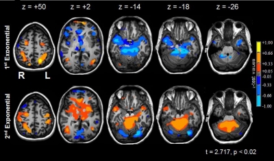
Fig. 5.
Changes in neural activity during sequence learning. Column 1 (z = +50): the significant activations during learning occurred within the left dorsal visual stream (i.e., left superior parietal lobule, L SPL) were confined to the 1st exponential period; bilateral dorsolateral premotor cortices (dlPMC) were active in both periods; primary motor cortex—hand area (L M1) was primarily active during the second exponential period. Column 2 (z = +2): the left medial head of the caudate nucleus (hCN) was below baseline in early learning and above baseline in late sequence learning; the bilateral frontal poles (FPs) were active in early learning and were below baseline in late sequence learning. Bilateral visual area 5/middle temporal areas (V5/MT) were active in both periods. Column 3 (z = −14): bilateral fusiform and lingual gyri (FUS/LING) were active in early learning and were below baseline in late sequence learning. Column 4 (z = −18): bilateral anterior vermis (aVER) was less active than baseline during early learning and more active than baseline in the 2nd exponential period. Column 5 (z = −26): the right dentate nucleus (DN) was less active in the 1st exponential period, relative to baseline, and was more active than baseline during late learning. See Table 4A for the percent signal changes and significance levels of selected regions of interest.

