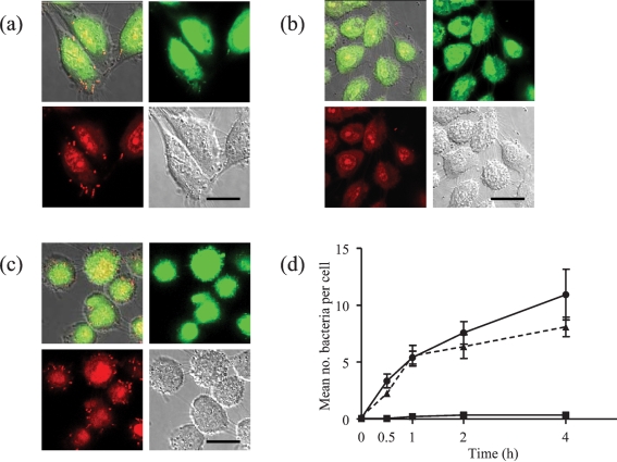
Fig. 1.
Interaction of S. gordonii with cultured oral keratinocytes after 2 h incubation. (a) Wild-type DL1, (b) srtA− mutant of DL1 and (c) complemented mutant (srtA+) were incubated with OFK6 TERT-2 oral keratinocytes in ibidi mini-flow cells for 2 h. After washing, adherent bacteria were stained using the BacLight Gram stain as described in Methods. Panels show the colour channel images superimposed upon differential interference contrast (DIC) images of the same cells, as well as each of the images (green, red and DIC) separately. Bars, 10 μm. (d) The mean number and se of bacteria per keratinocyte in each field for the wild-type (•), srtA− mutant (▴) and complemented mutant (▪) were calculated and plotted after 0, 0.5, 1, 2 and 4 h.

