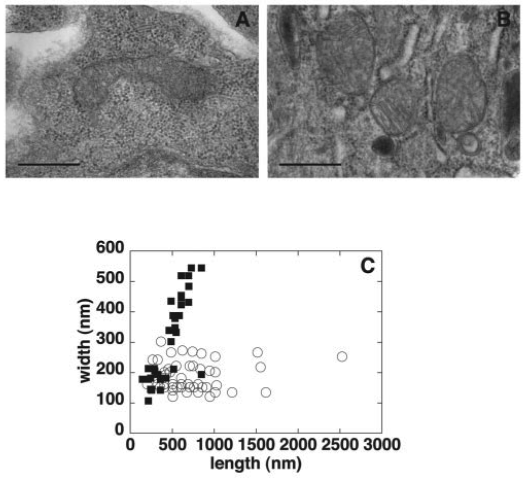
FIGURE 6. Electron microscopy of mitochondria in Pisd+/+ and Pisd−/− embryos.
Embryos (E8) were prepared for electron microscopy. Ultrathin sections were cut and stained with uranyl acetate and osmium tetroxide. A, representative elongated and tubular mitochondrion from a Pisd+/+ embryo. B, typical mitochondria from a Pisd−/− embryo; mitochondria are rounded and vesicular but cristae are visible. Size bar, 500 nm. C, length and width of mitochondria in Pisd+/+(open circles) and Pisd−/−(closed squares) embryonic tissue were measured from electron micrographs. Each symbol represents a single mitochondrion.

