
Figure 3.
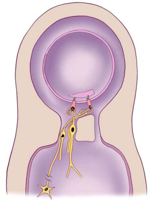
Simplified diagram showing the organization of inner ear organs of hearing and balance. The inner ear contains two fluid chambers, a membranous and a bony chamber. The membranous chamber is filled with endolymph while the bony chamber is filled with perilymph. The wall of the membranous chamber is made up of many cells that are so tightly joined together that they prevent the two fluids from mixing. The sensory epithelium makes up only a small portion of the wall of the membranous chamber and contains sensory receptor cells and surrounding supporting cells (supporting cells are not shown in the drawing).