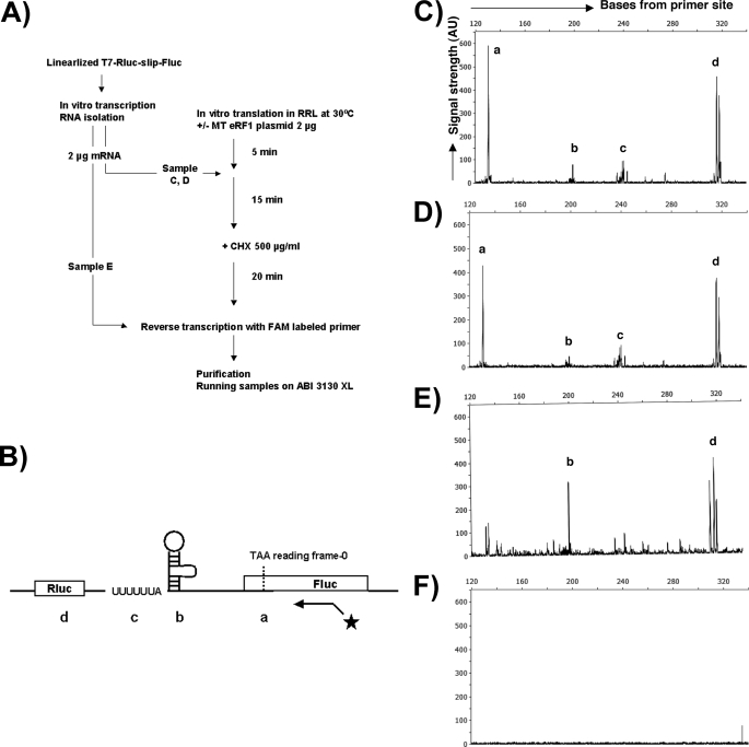
FIGURE 6.
Toeprinting indicates that mutant eRF1 does not promote ribosomal stacking at the PRF signal. A, flowchart of toeprinting assay. mRNAs transcribed from the linearized dual luc HIV-1 construct in vitro were added to rabbit reticulocyte lysates with or without the mutant eRF1 plasmid. After adding cycloheximide to arrest the ribosomes, mRNAs were reverse transcribed with a FAM-labeled primer downstream of the PRF signal. B, schematic representation of where peaks a, b, c, and d from panels C–E map including location of the primer. The starred line with arrow represents the binding position of the FAM-labeled primer. Peaks a, b, and c correspond to pausing at the in-frame stop codon, the stem and loop, and the slippery site, respectively. Peak d maps to a likely secondary structure in rluc. C, fragment analysis data from the sample with mutant eRF1. D, sample without mutant eRF1 plasmid. E, sample without incubation in rabbit reticulocyte lysate. F, no-mRNA control. RRL, rabbit reticulocyte lysate; MT, mutant; CHX, cycloheximide; AU, arbitrary unit.

