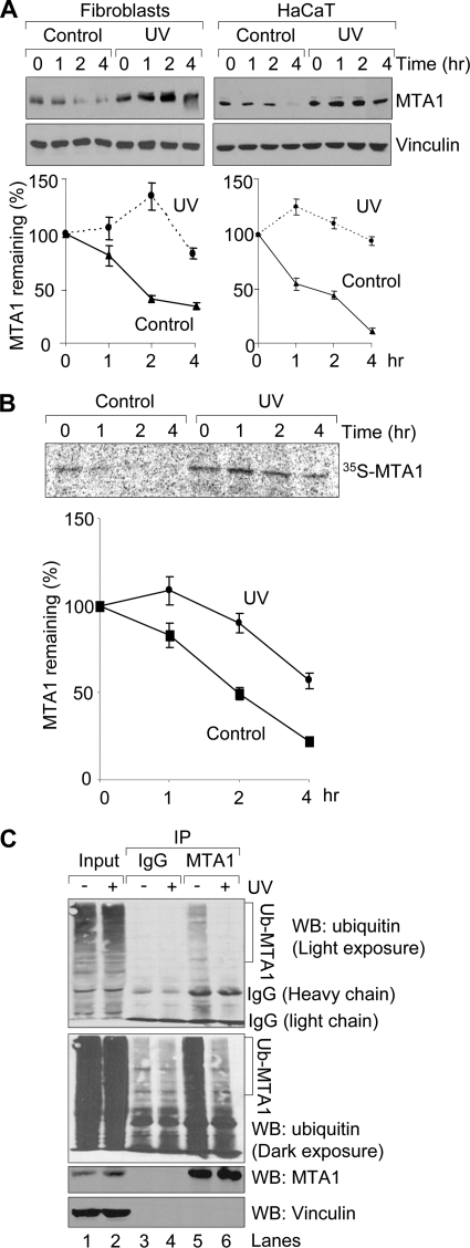
FIGURE 2.
UV stabilizes MTA1 by inhibiting its ubiquitination. A, HaCaT cells or fibroblasts were untreated or treated with 200 mJ/cm2 UV. After 1 h of UV treatment, cells were incubated with 100 μg/ml cycloheximide and harvested at the indicated time points for Western blotting analysis using the indicated antibodies. Western blots were subjected to densitometric analysis, and results were normalized based on vinculin expression levels and reported in a graph (bottom). The mean values from three independent experiments are shown. B, HaCaT cells were treated with 200 mJ/cm2 UV for 1 h prior to pulse-chase analysis using [35S]methionine labeling. Cells were harvested at various time points during the chase period and immunoprecipitated using an anti-MTA1 antibody. Complexes were resolved by SDS-PAGE and exposed to storage phosphor screens. The intensity of the labeled MTA1 band was quantified by PhosphorImager analysis using ImageQuant software (Amersham Biosciences), and the percentage of MTA1 remaining was calculated relative to that at the beginning of the chase period (time 0). The mean values from three independent experiments are shown. C, HaCaT cells were treated with 20 μm MG-132 for 1 h and then exposed to 200 mJ/cm2 UV. Protein extracts were prepared after 2 h of UV treatment and then subjected to sequential IP/Western blot (WB) analysis with the indicated antibodies.

