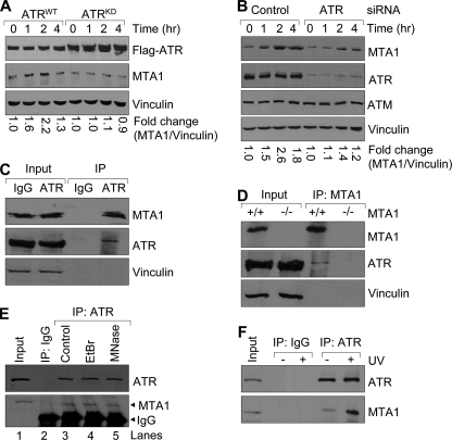
FIGURE 3.
Induction of MTA1 following exposure to UV is dependent on ATR. A, ATRWT- or ATRKD-expressing U2OS cells were incubated with 1 μg/ml doxycycline for 48 h and then exposed to 200 mJ/cm2 UV. Total cellular lysates were prepared at the indicated time points for Western blot analysis with the indicated antibodies. The density of bands was measured using the ImageQuest program and normalized to that of vinculin. The relative -fold change (MTA1/vinculin) is shown at the bottom. B, U2OS cells were transfected with specific siRNAs targeting human ATR or non-targeting control siRNAs. After 48 h of transfection, cells were treated with or without 200 mJ/cm2 of UV and then subjected to Western blot analysis with the indicated antibodies. The relative -fold change (MTA1/vinculin) is shown at the bottom. C, protein extracts from HeLa cells were subjected to IP analysis with an anti-ATR antibody or IgG control, followed by immunoblotting with the antibodies against ATR and MTA1. The immunoprecipitates were immunoblotted with vinculin, an abundant protein not expected to be part of this complex, as a negative control. D, protein extracts from MTA1+/+ or MTA1−/− (negative control) MEFs were subjected to IP analysis with an anti-MTA1 antibody, followed by immunoblotting with the indicated antibodies. E, HeLa cellular lysates were incubated with 100 μg/ml EtBr on ice for 30 min. Precipitates were removed by centrifugation for 5 min at 4 °C in a microcentrifuge, and the supernatant was transferred to a fresh tube. The resulting lysate was then ready for the IP assay. For micrococcal nuclease treatment, the immunoprecipitates bound to Protein G-agarose beads were incubated with micrococcal nuclease at 37 °C for 1 h, and the samples were washed twice with 1 ml of digestion buffer prior to SDS-PAGE. F, HeLa cells were irradiated with 200 mJ/cm2 UV and harvested after 2 h of UV treatment for sequential IP/Western blot analysis with the indicated antibodies.

