FIGURE 2.
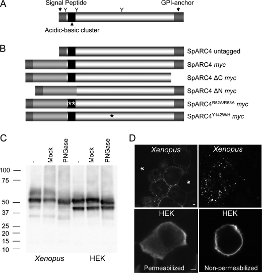
SpARC4 is a glycoprotein targeted to the plasma membrane. A, predicted structure of SpARC4. Putative N-linked glycosylation sites are marked with a Y. B, schematic of the constructs used in this study. C, Western blot analysis, using an anti-Myc antibody, of Xenopus embryos (left) or HEK cells (right) expressing Myc-tagged SpARC4 (SpARC4 Myc). Samples were untreated (−), mock-treated, or digested with PNGase-F prior to analysis. Migration of molecular mass markers (in kDa) is shown on the left side of the panel. D, confocal fluorescence images of X. laevis embryos (top) or HEK cells (bottom) expressing SpARC4 Myc. Expression was detected by immunocytochemistry using an anti-Myc primary antibody. For embryos, the images are typical of the two distribution patterns observed. The asterisks highlight cells not expressing the protein. For HEK cells, both permeabilized and nonpermeabilized cells were processed. Scale bar, 5 μm.

