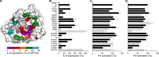
FIGURE 1.
Pro-coagulant and PAR2 signaling activities of FVIIa variants on human MDA-MB-231 breast cancer cells. A, structural model of the protease domain of FVIIa (surface representation) with the P5–P5′ fragment of PAR2 (sticks) bound in the active site. Positions in FVIIa evaluated by mutagenesis are color-coded according to the effect on PAR2-dependent IL-8 expression. B, IL-8 secretion was used as a measure of the PAR2-signaling response after stimulation of MDA-MB-231 cells with 10 nm FVIIa for 24 h. Variants with <25% or >200% of wt FVIIa-signaling activity are shown with light-colored bars. FVIIa Q40A and Q143N were indistinguishable from background levels (p > 0.05, unpaired t test). C and D, the pro-coagulant activity of each variant was measured on MDA-MB-231 cells with 10 nm FVIIa and 100 nm FX (C) or 200 nm FIX (D). Data are shown as mean ± S.D. (n = 3) normalized to wt FVIIa.

