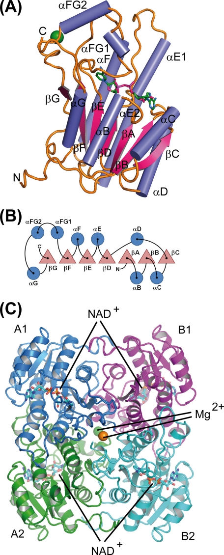
FIGURE 2.
Schematic representation of the secondary structure elements and their arrangement within GatDH. A, ribbon representation of one molecule of GatDH in complex with the cofactor NAD(H) (stick representation, carbon atoms colored in green) and one magnesium ion (green sphere). B, sketch of the arrangement of the secondary structure elements. Helices are represented by circles and β-strands by triangles. The nomenclature of secondary structure elements is according to 3α/20β-hydroxysteroid dehydrogenase (60). C, quaternary structure of GatDH. GatDH forms a homo-tetramer of point group symmetry D2. The individual protein chains are differently colored. Two magnesium ions (green and orange spheres) are coordinated each by two opposing C termini (A1–B2 and A2–B1, respectively). The active sites are positioned toward the surface of the protein as indicated by the NAD(H) molecules in ball-and-stick representation. The tetramer is oriented according to the PQR coordinate system (61) and displayed along the R-axis.

