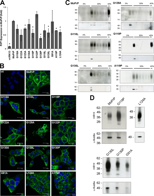
FIGURE 2.
GRR mutants are sorted normally. A, quantification of PrPC expression levels in cell lines; graph indicates normalized mean expression ± S.E. Asterisk indicates significant differences in expression (p < 0.05), and further details are available in supplemental Fig. S2. B, immunofluorescence of cell lines was performed with the PrP antibody ICSM-18 (green) and nuclear stain 4′,6-diamidino-2-phenylindole (blue). PrP was observed to localize to the plasma membrane and to an internal compartment consistent with the Golgi apparatus. This localization is similar in both MoPrP and GRR mutants, implying that mutations do not alter the subcellular expression of PrP. wt, wild type. C, cell lysates were subjected to limited Triton X-100 solubilization followed by sucrose density ultracentrifugation. Western blot analysis shows a concentration of MoPrP and GRR mutants at the interface of the 5 and 30% sucrose regions, where lipid rafts are expected to localize. The purification of lipid rafts was confirmed by probing for the raft-resident protein flotillin-2. D, exosomes were prepared from the media of transfected RK13 cells. Both prion-susceptible and prion-resistant cell lines show a concentration of PrP in exosomal pellets. Purification of exosomes was verified by probing for flotillin.

