FIGURE 3.
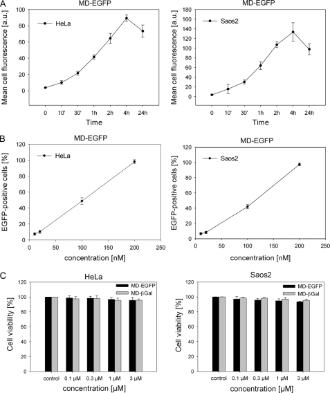
Kinetics of MD-EGFP internalization and cytotoxicity. A, time-dependent intracellular accumulation of MD-EGFP in HeLa and Saos2 cells. Both cell lines were incubated with 0.2 μm MD-EGFP at 37 °C for the times indicated and subsequently treated with trypsin for 10 min at 37 °C. The mean cell fluorescence was analyzed by flow cytometry. B, dose-dependent uptake of MD-EGFP in live HeLa and Saos2 cells. The indicated concentrations of MD-EGFP were added to the serum-free cell culture medium. After 4 h, cells were washed with PBS and trypsinized for 10 min at 37 °C, and cell fluorescence was measured by flow cytometry. All transduction experiments were performed in triplicates in two independent analyses, and the mean ± S.D. is indicated. C, MD-mediated protein delivery causes no cytotoxicity. HeLa and Saos2 cells were exposed to the indicated concentrations of MD-EGFP and MD-β-galactosidase (βGal) and incubated in growth medium at 37 °C for 24 h. Cytotoxicity after the uptake of both fusion proteins was determined by the leakage of lactate dehydrogenase into the culture medium. Each bar represents the mean ± S.D. of the viability of two independent experiments performed in triplicates. a.u., arbitrary units.

