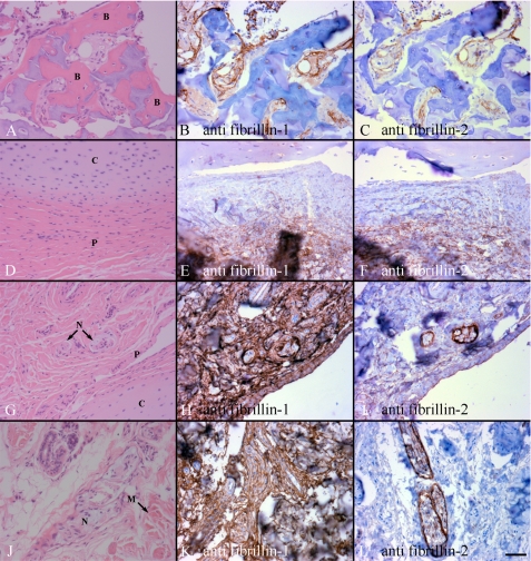
FIGURE 3.
Immunohistochemical staining of human postnatal supranummary digits. Sections were stained with mAb 15, specific for fibrillin-1 (B, E, H, and K), and with mAb 48, specific for fibrillin-2 (C, F, I, and L). Corresponding hematoxylin and eosin-stained sections are shown in A, D, G, and J. B and C, fibrillin-1 and -2 were found in the periosteum and marrow matrix around ossifying woven bone (labeled B in A) from a 17-month extra digit. E and F, staining for fibrillin-1 and -2 was found in the fibrous perichondrium next to the cartilage (labeled P and C in D) of a 17-month extra digit. H and I, fibrillin-1 was abundant, whereas fibrillin-2 appeared to be much less abundant, in all joint capsule tissues of a 21-month extra digit. Peripheral nerve (N), perichondrium (P), and cartilage (C) of the joint capsule are shown in G. K and L of 21-month digit, abundant fibrillin-1 staining was found around peripheral nerves and muscle (labeled N and M in J), whereas fibrillin-2 staining was abundant only around the nerves. Scale bar, 50 μm.

