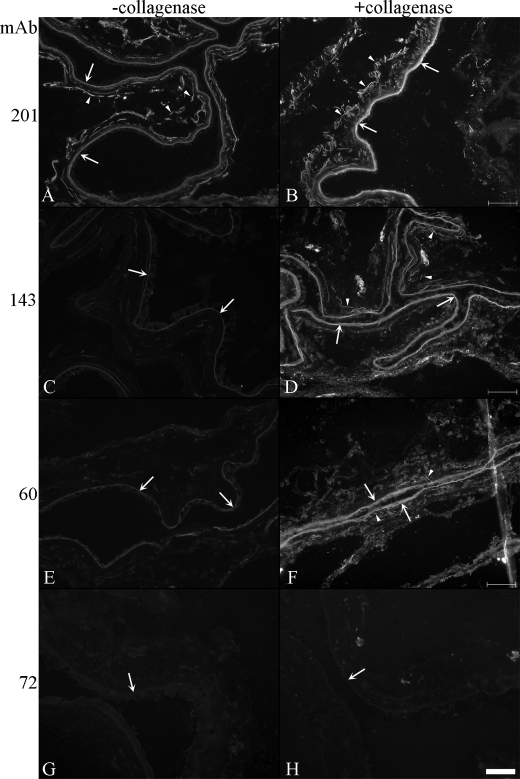
FIGURE 4.
Unmasking fibrillin-2 immunostaining in human amnion. Tissue sections in A, C, E, and G were not digested with crude collagenase, whereas those in B, D, F, and H were treated with enzyme. mAb 201, specific for fibrillin-1, stained microfibril bundles as well as the amniotic basement membrane in untreated sections (A) as well as collagenase-digested sections (B). mAb 143, one of two antibodies that recognize fibrillin-2 in fetal tissues, displayed only limited staining of the amniotic basement membrane in untreated sections (C). Digestion of tissue sections with crude collagenase unmasked the epitope recognized by mAb 143 in the microfibril bundles (D). mAb 60, which recognizes a cryptic epitope in fibrillin-2, gave similar results as mAb 143 (E and F). In contrast, mAb 72, a fibrillin-specific antibody, failed to recognize fibrillin-2 in both untreated (G) and treated (H) sections. This result was also obtained using mAb 205 (data not shown). Arrows point to the amniotic basement membrane; arrowheads point to microfibril bundles. Scale bar, 50 μm.

