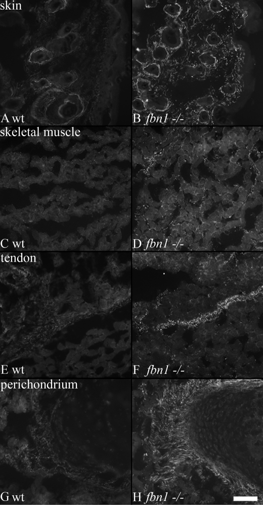
FIGURE 7.
Fibrillin-2 microfibrils are unmasked in mouse Fbn1 null tissues. P8 wild type (A, C, E, and G) and Fbn1−/− (B, D, F, and H) littermate sections were stained with pAb 0868, specific for fibrillin-2. Fibrillin-2 immunostaining was mostly negative in wild type sections of skin (A) and skeletal muscle (C) at this time; there was weak fibrillin-2 immunostaining in wild type sections of tendon (E) and perichondrium (F). However, in Fbn1−/− sections, fibrillin-2 immunostaining was revealed in typical tissue-specific fibrillin microfibril patterns (B, D, F, and H). Scale bar, 50 μm.

