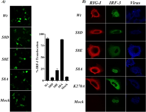
FIGURE 3.
Functional effect of RIG-I serine 8 substitutions on IRF-3 localization. A, GFP-IRF-3 localization in 293T cells co-transfected with RIG-I CARD WT or with the indicated serine 8 mutants. Images of live cells were taken 12 h after transfection, and translocation percentage was calculated by dividing the amount of cells with nuclear GFP-IRF3 by the total number of GFP-positive cells. Left, live image of GFP fluorescence in co-transfected cells. Right, quantification of nucleus/cytoplasm ratio (translocation) of GFP-IRF-3. Three individual transfection experiments for each CARD construct and four fields for each transfection experiment were used for the analysis. Error bars represent the S.D. for the different measurements. B, immunofluorescence for overexpressed full-length RIG-I WT or mutants (red) or GFP-IRF-3 (green) in HeLa cells infected with influenza A/PR/8/34 ΔNS1 virus (blue).

