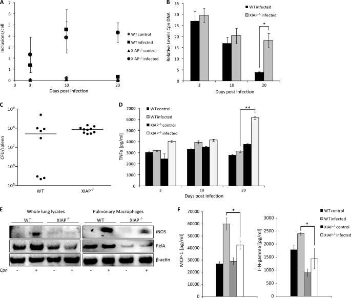
FIGURE 1.
Sensitization of XIAP KO mice for C. pneumoniae lung infection. A, impaired resolution of C. pneumoniae infection from the lungs of XIAP deficient mice is shown. Five mice per group were infected with C. pneumoniae, and infectious bacteria were determined by lung infectivity assays using quantitative immunofluorescence microscopy. The infectivity was calculated as the number of chlamydial inclusions formed per infected HEp2 cell. The data are represented as the mean ± S.E. of the inclusions per cell from three independent experiments. B, five mice per group were infected with C. pneumoniae, and the chlamydial ompA gene was amplified from the lung tissue of each mouse. The relative amount of the ompA gene in each lung sample was calculated using glyceraldehyde-3-phosphate dehydrogenase as an internal control. *, p ≤ 0.05. C, shown is the bacterial load after intravenous infection with S. typhimurium. C57BL/6 and KO XIAP mice were infected with 500 cfu of S. enterica typhimurium SL1344 via tail vein. cfu per spleen were determined 5 days post-infection. Bacterial load is equivalent in both C57BL/6 and KO XIAP mice. D, increased TNF-α production in lungs of infected XIAP KO mice is shown. Levels of TNF-α were determined in lung homogenates from WT and XIAP-deficient mice by enzyme-linked immunosorbent assay at different time points post infection. Data represent the means ± S.E. from five independent infection experiments. ** indicates p ≤ 0.01. E, reduced expression of iNOS and RelA/p65 in infected XIAP KO mice is shown. Lungs were excised aseptically from WT and XIAP KO mice infected with C. pneumoniae (Cpn) 20 days. CD11b+ pulmonary macrophages were purified from the lungs of WT and XIAP KO mice as described under “Experimental Procedures.” Homogenized lungs and isolated macrophages were lysed in radioimmune precipitation assay buffer containing protease inhibitors. 20 μg of protein from either whole lung lysate or pulmonary macrophages was separated by PAGE and subjected to Western blot analysis as described under “Experimental Procedures.” β-Actin was detected as the loading control. F, reduced secretion of major macrophage stimulatory cytokines is shown. The lung homogenate from mice of different groups were prepared, and the titers of IFN and MCP-1 were quantified as described under “Experimental Procedures.” Data represent the means ± S.E. from three independent infection experiments. * indicates p ≤ 0.05.

