FIGURE 5.
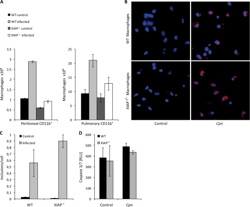
Depletion of macrophages in infected XIAP KO mice. A, shown is depletion of mouse peritoneal macrophages in infected XIAP KO mice. Peritoneal macrophages were isolated from different experimental groups, and their viability was tested by trypan blue staining. Data represent the mean of the number of viable macrophages ± S.E. from three independent experiments. B, infection of macrophages by C. pneumoniae is shown. Peritoneal macrophages from wild type and KO mice were infected with C. pneumoniae at a multiplicity of infection of 1. 72 h post-infection, the macrophages were fixed with chilled absolute methanol and stained with anti C. pneumoniae (Cpn) antibody (red) and Hoechst to stain for nuclei (blue). Shown are representative micrographs of infected macrophages. C, shown is quantification of the experiment shown in B. D, C. pneumoniae infection does not activate caspase-3 in XIAP KO Mac-1+ peritoneal macrophages. 104 macrophages from WT and XIAP KO mice were infected and co-stimulated with TNF-α and IFN-γ as indicated. Caspase-3 activity was quantified 24 h post-infection by luminescence assay. Data are represented as the mean ± S.E. from three independent experiments.

