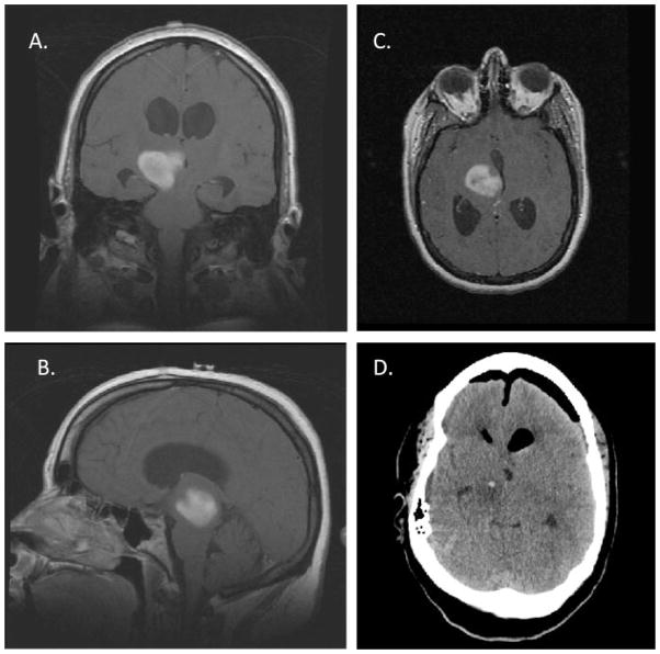
FIGURE 6.
Imaging of a 23–Year–Old Female Who Underwent a Neuroendoscopic Biopsy of a Thalamic Malignant Glioma and an Endoscopic Third Ventriculostomy for Treatment of Her Obstructive Hydrocephalus. (A) Coronal, (B) sagittal, and (C) axial T1-weighted magnetic resonance imaging of the brain after gadolinium administration revealed contrast enhancement of a right thalamic mass (indicated by arrows) extending into the midbrain and causing obstructive hydrocephalus. (D) A postoperative computed tomography scan of the brain demonstrating ventricular decompression and an area of neuroendoscopic sampling of the tumor (indicated by arrow) was obtained for pathologic diagnosis.

