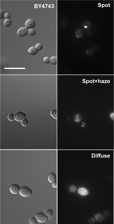
Figure1. The localization of Gsy2-GFP within wild type yeast cells.
The wild type yeast strain BY4743 was transformed with a centromeric plasmid encoding the GSY2 open reading frame fused at the C-terminus to GFP (pJR1420-A). After overnight growth in SC-Ura medium, aliquots of cells were examined by fluorescence microscopy. The left-hand series of images shows representative fields of cells observed using differential interference contrast. The right-hand series of panels shows fluorescence images of the same fields. Three distinct types of localization pattern could be distinguished within a single cell population; well defined bright spots (spot), spots accompanied with a haze (spot+haze), and diffuse cytoplasmic staining (diffuse). The scale bar represents 10 μm.

