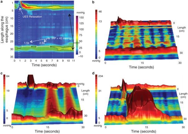
Figure 3.
Achalasia subtypes are distinguished by three distinct manometric patterns of esophageal body contractility. In classic achalasia (panel a as isobaric contour plot and panel b as a landscape plot), there is no significant pressurization within the body of the esophagus and impaired EGJ relaxation (IRP of 42 mm Hg in this example). The black 42 mm Hg isobaric contour line isolates the portions of the EGJ and UES recording during which the pressure is greater than 42 mm Hg. Panel c represents a swallow from a patient the “achalasia with compression” subtype exhibiting pan-esophageal pressurization. Although high pressures may be recorded in the esophageal body in this subtype, pressure is generated by esophageal shortening in conjunction with contraction of both sphincters, rather than by spasm in the esophageal body. Radiographically, there is no esophageal retention in these patients. Panel d illustrates a pressure topography plot illustrative of spastic achalasia. The three-dimensional rendering of panel d highlights the peaks and valleys of that spastic contraction that would likely appear as a corkscrew pattern on fluoroscopy. These patients often have diffuse thickening of the distal esophageal muscularis propria. EGJ, esophagogastric junction; IRP, integrated relaxation pressure.

