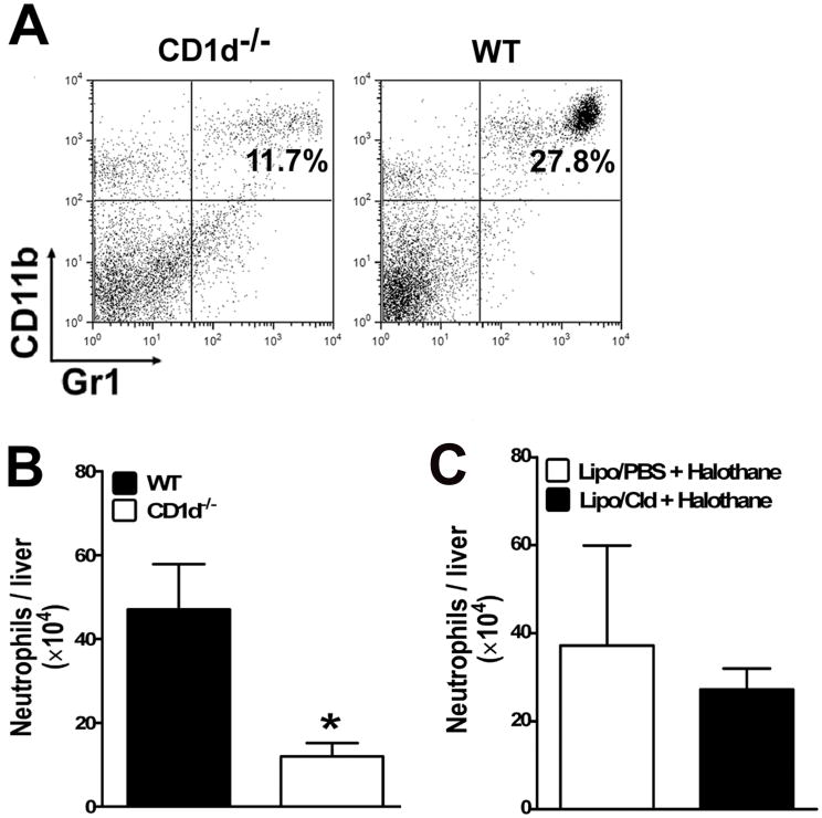
Figure 6.
Halothane-induced hepatic recruitment of neutrophils was not affected by KC depletion, but was inhibited in CD1d−/− mice compared with WT mice. (A and B) Female WT and CD1d−/− mice were treated with halothane. Hepatic leukocytes were isolated 12 h later from individual mice and stained with FITC-conjugated anti-Gr-1 and PE-conjugated anti-CD11b antibodies and analyzed by flow cytometry. (A) Data shown are representative dot plots of neutrophil infiltration into the liver of halothane-treated WT and CD1d−/− mice. The percentage of neutrophils (Gr-1+CD11b+) is presented in the upper right quadrant of each graph. (B) The absolute number of infiltrated neutrophils was calculated as the product of the total number of hepatic leukocytes isolated and the percentage of neutrophils. The absolute number of hepatic neutrophils per liver from halothane-treated WT and CD1d−/− mice (10 mice per group) are plotted as mean ± SEM. (*, p <0.05 compared with WT mice). (C) KCs were depleted as described in Fig. 3, and treated with halothane 2 days later. Hepatic leukocytes were isolated 12 h after halothane treatment, and the cells were stained and analyzed by flow cytometry as described above. The absolute number of infiltrated neutrophils per liver were calculated and plotted as mean ± SEM.

