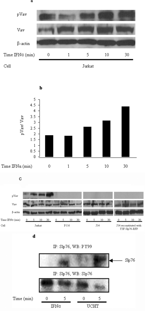
Figure 2. Activation of both Vav and Slp76 by IFNα mirrors the TCR ESC response in Jurkat and primary T-cells.
(a) A Western blot was performed to determine the expression levels of phospho(Tyr174)-Vav and Vav in IFNα-stimulated Jurkat cells over the indicated time course. β-Actin was used as a loading control. (b) Phospho(Tyr174)-Vav levels normalized to Vav expression by densitometric analysis. (c) Western blotting was performed to determine the expression levels of phospho(Tyr174)-Vav and Vav in IFNα-stimulated Jurkat, Zap70-deficient P116 cells and Slp76-deficient J14 cells over the time periods indicated. β-actin was used as a loading control. (d) Immunoprecipitation (IP) of Vav and Slp76 from primary T-cell lysate. Probing with anti-phosphotyrosine antibody (pY99) reveals that Slp76 and Vav are tyrosine-phosphorylated following 5 min of IFNα stimulation. As a control, the TCR was also stimulated for the same time period and the UCHT1 antibody was used to show the expected protein phosphorylation.

