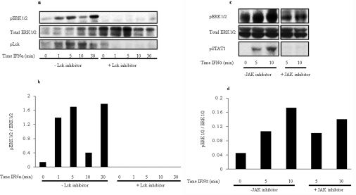
Figure 5. ERK is phosphorylated in response to IFNα in primary human CD4+ T-cells: components of the TCR machinery are required for this ERK phosophorylation.
(a) Freshly isolated human peripheral CD4+ T-cells were stimulated for up to 30 min in the presence and absence of Lck inhibitor I. UCHT1 was used to stimulate cells as a control for 5 min in the presence and absence of inhibitor. Lysates were immunoblotted with anti-phospho-ERK1/2 antibody (pERK1/2; top panel), anti-phospho-Lck antibody (pLck; bottom panel) or anti-ERK1/2 antibody (total ERK 1/2; middle panel). (b) Normalized densitometric analysis of ERK phosphorylation. The level of increased ERK activity is relative to the level of total protein. (c) Human peripheral CD4+ T-cells were stimulated for the time periods indicated without (left-hand side) or with 15 nm (right-hand side) of JAK-1 inhibitor for 5 and 10 min on the same gel. The same basal time point (no IFNα added) was used, as shown in the left-most lane. Lysates were used to immunoblot with either anti-phospho-ERK1/2 antibody (top panel), anti-phospho-STAT1 antibody (bottom panel) or anti-ERK1/2 antibody (total ERK 1/2; middle panel). (d) Densitometric analysis of ERK phosphorylation taking into account level of protein present in each lane. The level of ERK phosphorylation is shown in arbitrary units.

