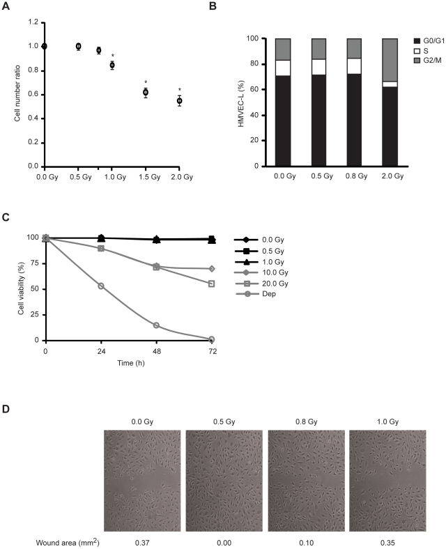
Figure 1. Low-dose IR promotes endothelial cell migration without causing cell cycle arrest or apoptosis.
(A) HMVEC-L were plated at equal densities and, after 12 h, left untreated or exposed to 0.5, 0.8, 1.0, 1.5 and 2.0 Gy. After 72 h, the cells were counted using a Nucleocounter. The values (means ± s.d.) represent the ratio between cell number of irradiated and non-irradiated conditions and are derived from four independent experiments. * P<0.02. (B) HMVEC-L were exposed or not to 0.5, 0.8 and 2.0 Gy. Cell cycle profiles were assessed by flow cytometric analysis after 72 h of culture. Data are representative of three independent experiments. (C) The percentage of apoptotic cells was determined by flow cytometry at the indicated time. Cells cultured without serum (Dep) were used as cell death control. Values are given as the percentage of viable cells (Annexin V, PI negative) remaining in culture. Data are shown as mean in triplicate culture and are representative of three independent experiments. (D) Confluent monolayers of HMVEC-L were subjected to in vitro wound healing and exposed or not to 0.5, 0.8 or 1.0 Gy. Photographs were taken immediately (not shown) and 9 h after wounding. Quantification of the wound area (in mm2) is presented below the images. Data are representative of five independent experiments.

