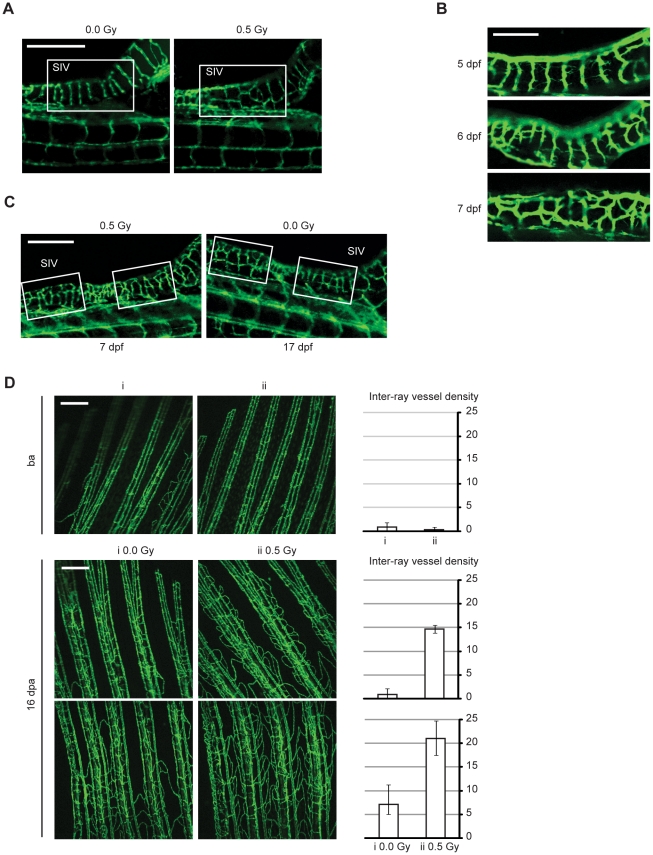
Figure 5. Low-dose IR accelerates angiogenic sprouting during zebrafish embryonic development and enhances angiogenesis during fin regeneration.
(A–C) Live zebrafish embryos were exposed or not to 0.5 Gy IR 3 d post-fertilization (dpf). Representative images of Sub-Intestinal Vessels (SIV), from (A) a non-irradiated and irradiated zebrafish at 7 dpf; (B) an irradiated zebrafish at 5 (top), 6 (middle) and 7 dpf (bottom); (C) an irradiated zebrafish at 7 dpf and a non-irradiated zebrafish at 17 dpf. Scale bars, 250 µm (A and C), 100 µm (B). (D) Fli1:EGFP adult zebrafish caudal fins were amputated at mid-fin level, exposed or not to 0.5 Gy of IR and then allowed to recover. Representative images from vasculature of two zebrafish fins (i and ii) before amputation ensure they had identical vasculature (top). Representative images from vasculature of two different fin areas of the same zebrafish, 16 days post-amputation (dpa), with or without low-dose IR treatment (middle and bottom). Each image was quantified for inter-ray vessel density. Data are shown as mean and error bars indicate maximum and minimum values. Images are representative of 10 zebrafish in five independent experiments. Scale bars, 250 µm.

