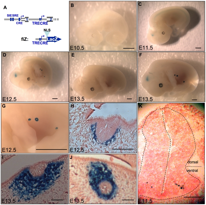
Figure 4. Transgenic analysis of the putative promoter shows expression restricted to the spinal cord and mammary bud of mouse embryos.
A. 5′ part of mouse c-fos gene (up) and our NLS-containing, betagalactosidase reporter construct (bottom), fiZ (fos intron lacZ). 7 Transgenic mouse lines were created with fiZ and transgenic embryos were stained for betagalactosidase activity. Here are shown transgenic embryos from mouse line #60 at day 10.5 (B), 11.5 (C, K), 12.5 (D, G, H), and 13.5 (F, I, J). B, C, D, F: whole-mount embryos showing the spinal cord staining starting day 11.5 p. c. and the mammary gland anlage staining starting 12.5 p. c. G. Close-up view of the developing mammary buds showing the stained ring corresponding to mesenchymal cells. H, I, J: sagittal frozen sections showing the nuclear staining in the mesenchymal part of the mammary bud, but not in the central, epithelial part. K. Transverse frozen section on a 11.5 d. p. c. embryo showing the extremely restricted, ventral spinal cord staining. E: wild-type, e13.5 embryo as a control. Scale bars: 1 mm (B, C, D, E, F, G) then 150 µm (H, I, J, K).

