
Figure 1.
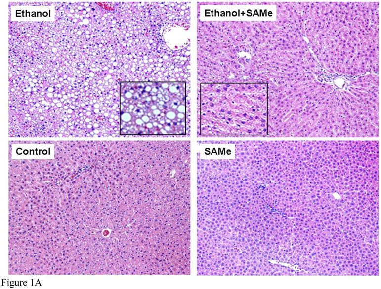

Figure 1A. Liver sections were stained with hematoxylin and eosin. Hepatocytes from rats fed ethanol only showed macrovesicular steatosis (x130). Hepatocytes from rats fed ethanol plus SAMe showed microvesicular steatosis (x130). Magnification of inserts was x 260. Hepatocytes from rats fed dextrose showed normal liver without steatosis (x130). Hepatocytes from rats fed SAMe alone, showed normal liver without steatosis (x130)
Figure 1B. Morphometric quantitation of liver fat. Ethanol fed alone caused a marked increase in fat, significantly greater than the 3 other groups of rats (Mean±SEM n=3 p<0.05). Ethanol fed with SAMe caused less steatosis but significantly more than the two controls (Mean±SEM, n=3, p<0.05).