
Figure 1.
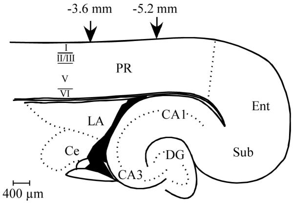
Schematic diagram of rat perirhinal cortex. A horizontal brain slice containing rat perirhinal cortex (PR) with the relative laminar boarders indicated on the left side. Neuronal counting and digital analyses were conducted within the rostro-caudal boundaries of PR indicated by the downward arrows. Abbreviations: CA1, area CA1 of the hippocampal formation; CA3, area CA3 of the hippocampal formation; Ce, Central Nucleus of the Amygdala; DG, Dentate Gyrus; LA, lateral nucleus of the amygdala; Ent, entorhinal cortex; PR, perirhinal cortex; Sub, subiculum.