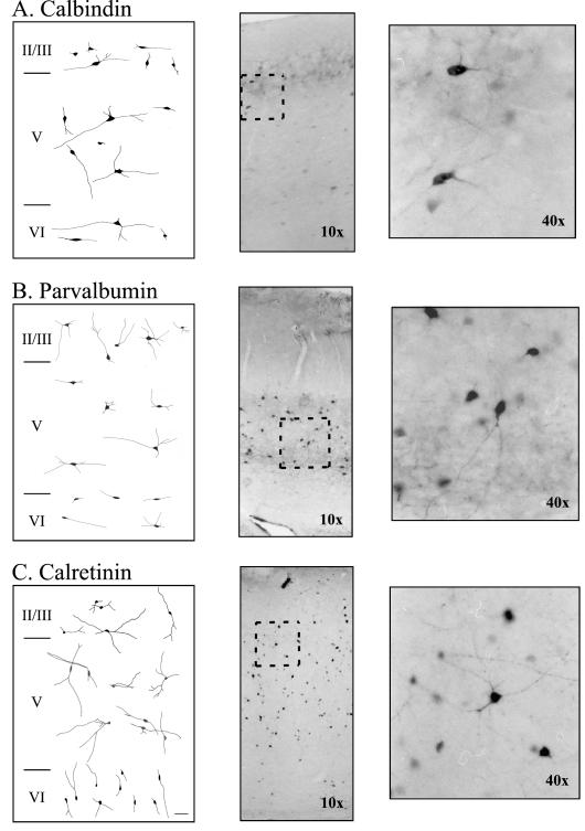
Figure 3.
Morphology of neurons immunoreactive for calcium binding proteins in adult rat perirhinal cortex. (A-C) Representative neuronal morphologies of calbindin-, parvalbumin, and calretinin-immunoreactive neurons in perirhinal layers II/III, V, and VI. The left panel shows camera lucida reconstructions, the middle panel shows a representative photograph using a 10X air objective. The area inside the rectangle is magnified (using a 40X air objective) and shown in the right panel.

