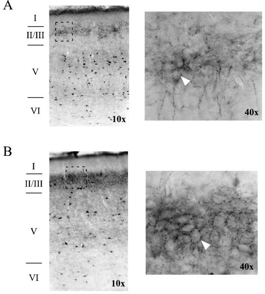
Figure 4.
Axons immunoreactive for parvalbumin form baskets around unstained cell bodies in rat perirhinal cortex. (A) Horizontal section from an adult animal taken at −7.6 mm D/V relative to Bregma. On the left is an image from a 10X air objective, showing all layers of PR. The area in the rectangle (in layer II/III) is shown on the right using a 40X air objective. The arrowhead points to the outline of a cell body. (B) Horizontal section from a middle-aged animal taken at −7.34 relative to Bregma. Images on the left and right, respectively, were taken using 10X and 40X air objectives. The arrowhead points to the outline of a cell body.

