
Figure 4. Neuronal excitability is comparable in CA1 hippocampal neurons from 2 mo old WT and 5XFAD mice.
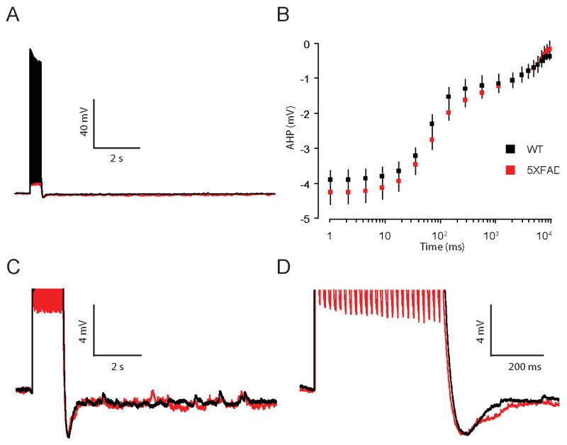
A, Representative traces of the AHP measured in whole-cell current clamp mode from neurons of 2 mo old 5XFAD (red) and WT (black) mice. AHPs were elicited by 25 brief current injections to elicit 25 action potentials at 50 Hz. B, Summary plot of the AHP versus time on a log scale for 2 mo old 5XFAD (red) and WT (black) mice. The AHP was averaged into bins to give mean and SEM at multiple time points (see Methods). No significant differences in the mean AHP were observed in 5XFAD compared to WT mice at 2 mo of age (p = 0.4). C,D, AHP on an expanded scale.