Figure 3.
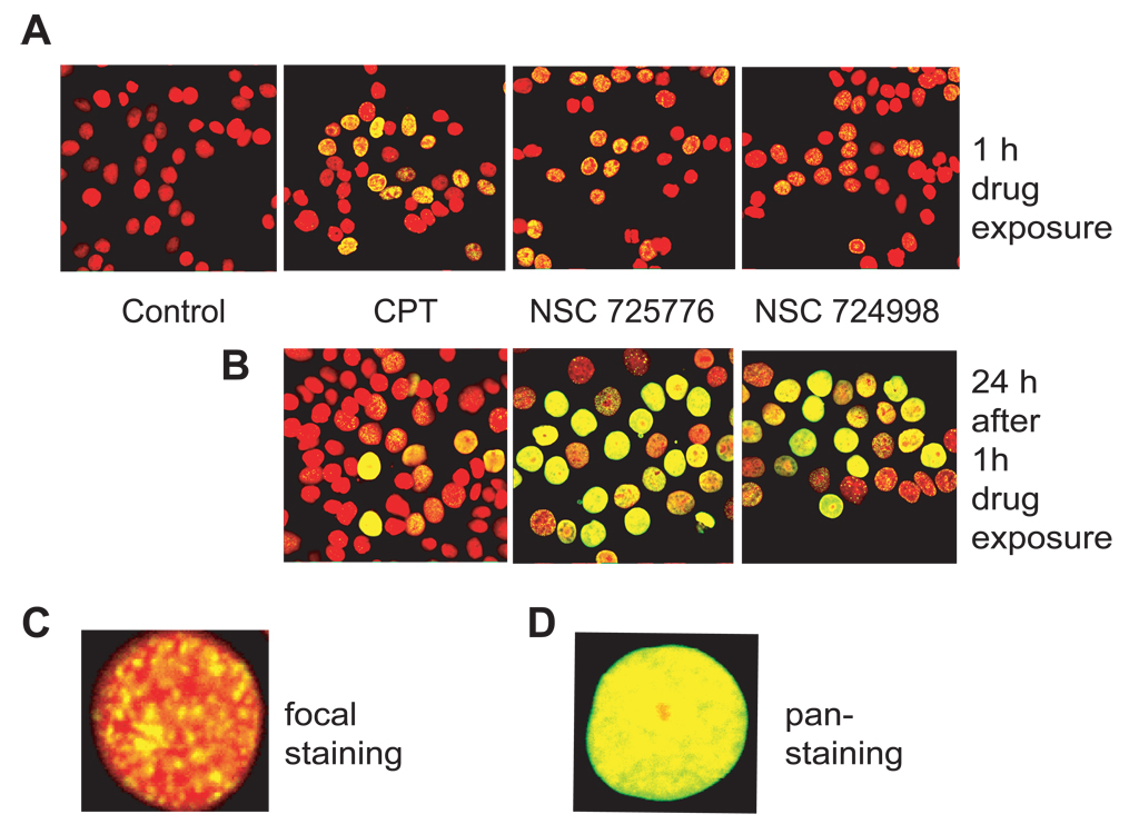
γ-H2AX induction by NSC 725776 and NSC 724998 in human colon cancer HT29 cells. A) Rapid induction of γ-H2AX foci after 1 h treatment with 1 µM of CPT, NSC 725776 or NSC 724998. B) Persistent γ-H2AX signal induced by indenoisoquinolines. After 1 h drug treatments, HT29 cells were further incubated in drug-free medium for 24 h. C) Representative image of a single cell with typical γ-H2AX damage foci. D) Representative image of a single cell with diffuse pan-nuclear staining, which typically develops several hours after drug exposure. Fixed cells were stained with mouse anti-γ-H2AX antibody and goat anti-mouse antibody conjugated with AlexaFluor 488 (green). Nuclei were stained with PI (red).

