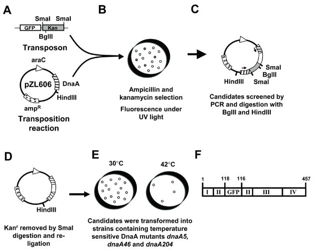
Figure 1.
Schematic of gfp-dnaA Construction (A–F). (A) The transposons were amplified and moved into DnaA expression plasmid pZL606 by a transposition reaction. (B) Candidate colonies were selected by ampicillin and kanamycin resistance and for fluorescence under UV light illumination. (C) Candidates were then screened for GFP insertion by digestion with BglII and HindIII and by PCR. Arrows denote primer locations. (D) KanR was removed by SmaI digestion and the plasmid was re-ligated. (E) Candidates were selected and screened for DnaA function by rescue experiments with DnaA temperature-sensitive mutants. F) Schematic of the insertion of GFP into DnaA in candidate plasmid pYYH327. GFP follows residue 118 of DnaA, and at the carboxy end of GFP, DnaA resumes beginning with residue 116. This candidate showed the best results in the temperature sensitive rescue assays and was selected for allelic replacement and use in the remainder of this paper.

