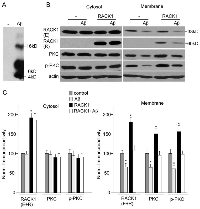
FIG. 1.
Aβ42 treatment decreases RACK1, total and activated PKC in membrane fractions of cultured cortical neurons. A, Western blots showing the oligomeric Aβ B, Western blots showing endogenous (E) and recombinant (R) RACK1, PKC, p-PKC and actin in cytosolic and membrane fractions from cortical cultures (uninfected or infected with GFP-RACK1 virus) treated without or with Aβ oligomer (1 μM, 48 hrs). C, Quantifications by densitometric analysis of RACK1 (E+R), PKC, p-PKC in cytosolic and membrane fractions from non-treated or Aβ-treated cortical cultures without or with RACK1 overexpression. Protein levels (normalized to actin) were expressed as the percent of controls. *: p < 0.01, ANOVA, compared to untreated and uninfected neurons (control).

