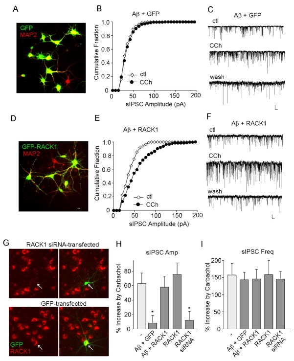
FIG. 3.
Overexpression of RACK1 restores the Aβ-induced loss of muscarinic regulation of sIPSC amplitudes in cortical cultures. A,D, Immunocytochemical images of MAP2(red)-stained cortical cultures infected with GFP (A) or GFP-RACK1 (D) Sindbis viruses. B,E, Cumulative plots of the distribution of sIPSC amplitudes showing the effect of carbachol (CCh, 20 μM) in A (1 μM, 2–3 days)-treated cortical cultures infected with GFP (B) or GFP-RACK1 (E) Sindbis viruses. C,F, Representative sIPSC traces from the neurons used to construct B and E. Scale bars: 30 pA, 1s. G, Immunostaining of RACK1 (red) in cultured cortical neurons transfected with RACK1 siRNA (co-transfected with GFP) or GFP alone. Arrowheads point to GFP+ neurons. H, I, Cumulative data (mean ± SEM) showing the percent increase of sIPSC amplitude (H) or frequency (I) by carbachol in cortical neurons under different conditions. *: p < 0.01, ANOVA, compared to the effect of carbachol in the control condition (−).

