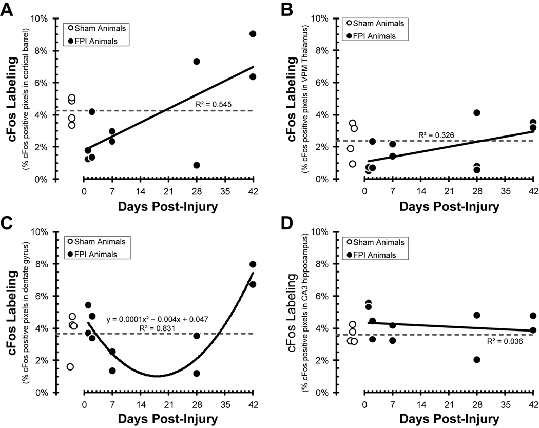
Figure 4.
Pixel quantification of cFos labeling of uninjured (open circles) and fluid percussion injured (FPI) animals (closed circles) over a 42 day post-injury time course (see methods for details). The open circles and horizontal dashed line indicate activation following whisker stimulation in individual uninjured sham animals and the group average, respectively. (A) In the somatosensory barrel fields, over the first week post-injury, fewer cortical barrel pixels were activated by whisker stimulation. At 28–42 days post-injury, cFos labeling exceeds sham levels. The positive correlation between cFos labeling and time post-injury (r2=0.545) indicates a greater extent of neuronal activation following diffuse brain injury. (B) In the ventral posterior thalamus barreloids, over the first week post-injury, fewer pixels were activated by whisker stimulation. At 28–42 days post-injury, cFos labeling returns to sham levels. (C) In the hippocampus dentate gyrus, cFos labeling declined over the first week post-injury, remained depressed through 28 days, and exceeded sham values by 42 days post-injury. The data fit a U-shaped, second-order polynomial correlation between time post-injury and cFos labeling (r2= 0.831). (D) In the hippocampus area CA3, cFos labeling remained unchanged from sham values over the 42 day time course.

