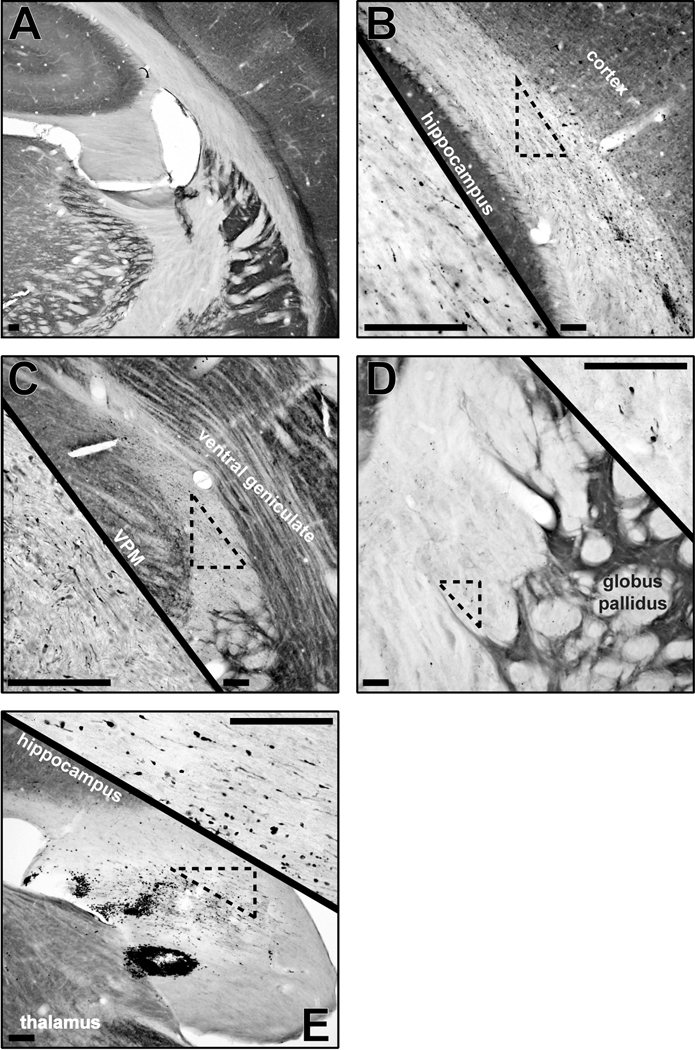
Figure 5.
Cytochrome oxidase histochemistry (background stain) with amyloid precursor protein (APP) immunohistochemistry (black) in uninjured sham (A; −3.24 mm from bregma) and 2d brain-injured (B–E) rats demonstrate the extent of axonal pathology across the subcortical white matter (B; −4.56 mm from bregma), superior thalamic radiation (C; −4.56 mm from bregma), internal capsule (D; −1.56 mm from bregma) and fimbria of the hippocampus (E; −3.48 mm from bregma). Axonal injury is identified by APP-positive immunoreactivity and swollen axonal segments or terminal bulbs. Scattered pathology is evident in the low magnification images and the high magnification insets. All scale bars are 10 µm.

