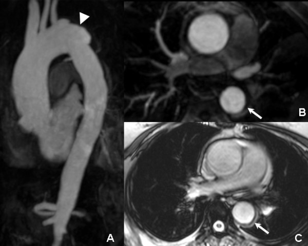
Figure 3.

Three months follow-up CMR: the MIP (A), MPR (B), and true FISP axial images (C) revealed partial regression of the hematoma (arrow) and complete absorption of the pleural effusion, while the ulcer-projection lesion progressed into an aneurysm like contour (arrowhead).
