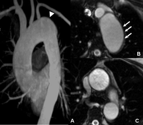
Figure 4.

One year follow-up CMR: the MIP (A), MPR (B), and true FISP axial images (C) revealed that the aortic arch aneurysm (arrowhead) was larger than it was at the 3 months follow-up examination. A thickened aortic wall without an apparent low signal non-enhanced hematoma could be seen, which suggested the IMH was reabsorbed.
