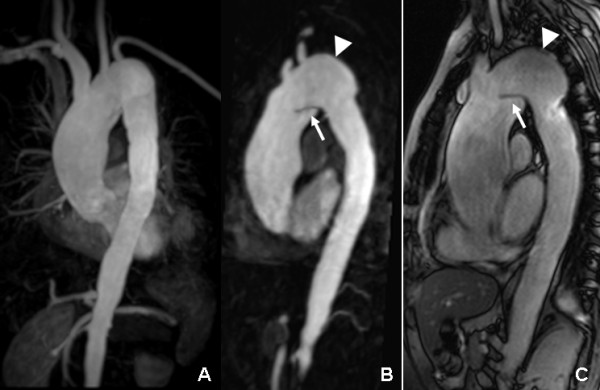
Figure 5.

Six year follow-up CMR: the MIP (A) image showed the extended widening lumen of ascending aorta and aortic arch with irregular wall. The MPR (B) and true FISP sagittal (C) images displayed the luminal intimal flap (arrow) located at aortic arch that formed limited aortic dissection. The aortic arch aneurysm was still present (arrowhead).
