
Figure 1.
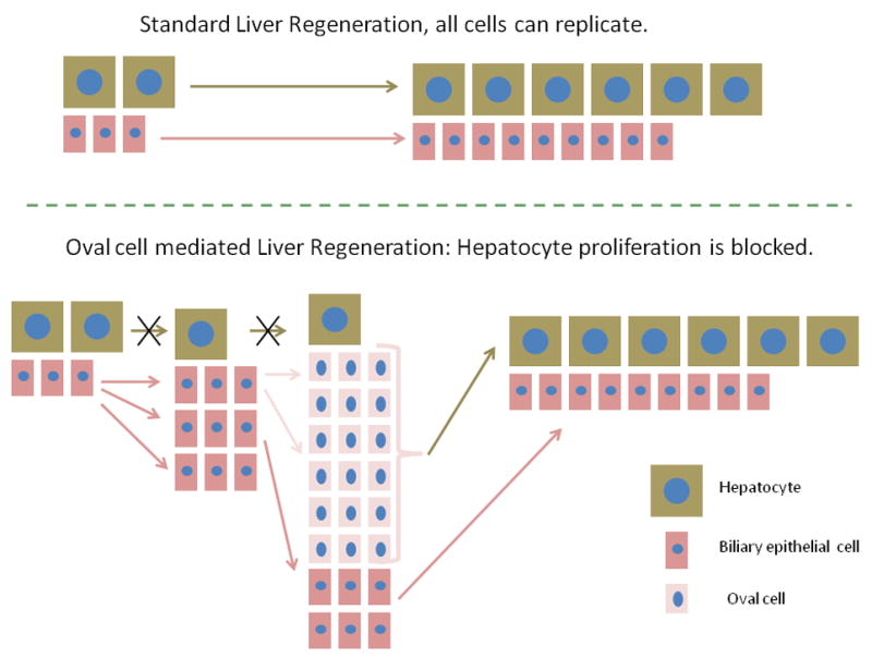
In standard liver regeneration, as after 2/3 partial hepatectomy, all cells are capable of proliferation. Hepatocytes give rise to hepatocytes and biliary cells generate biliary cells. When hepatocyte proliferation is blocked, biliary cells replicate themselves and they also expand into a large number of oval cells, with gene expression patterns of both biliary and hepatocyte specificity. Sources of oval cells are portal ductules and biliary cells of the canals of Hering. The oval cells transition into a hepatocyte phenotype and rescue the hepatocyte compartment.