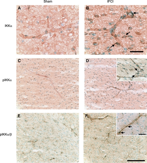
Figure 3.
Immunohistochemistry of IKKα, pIKKα, and pIKKα/β 1 h after 30 mins of tFCI in mice. (A) Weak IKKα immunopositivity was observed in sham-operated mice. (B) Cerebral ischemia induced an increase in expression and nuclear accumulation of IKKα in endothelial cells of the infarcted brains. (C, E) Phosphorylation of IKKα and IKKα/β was detected at low levels in sham-operated mice. (D, F) After tFCI, pIKKα and pIKKα/β significantly increased in endothelial cells 1 h after 30 mins of tFCI. Scale bar=25 μm (panel B), scale bar=100 μm (panel F), and scale bar=25 μm (panel F, inset).

