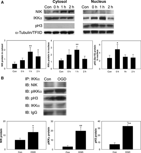
Figure 4.
Changes in NIK, IKKα, and pH3 (Ser10) after 4 h of OGD in mouse cerebral endothelial (bEnd.3) cells. (A) NIK, an upstream kinase of IKKα, and pH3 (Ser10) were increased compared with the controls (Con) in accordance with nuclear accumulation of IKK in bEnd.3 cells. α-Tubulin and TFIID were used as internal controls. Quantitative analysis showed the relative changes in the amount of NIK, IKKα, and pH3 (Ser10) (n=4, *P<0.05, **P<0.01 compared with controls). (B) Endogenous IKKα was captured by immunoprecipitation (IP) with an anti-IKKα antibody in the total fraction of bEnd.3 cells. Results were detected by immunoblotting (IB) using antibodies to NIK, pIKKα, and pH3. Coimmunoprecipitation analysis for pIKKα/NIK/pH3 in bEnd.3 cells showed significant increases in OGD in bEnd.3 cells compared with control cells. Input lysates showed no differences. Grouped quantitative data are presented as mean±s.d. (n=3, *P<0.05, **P<0.01).

