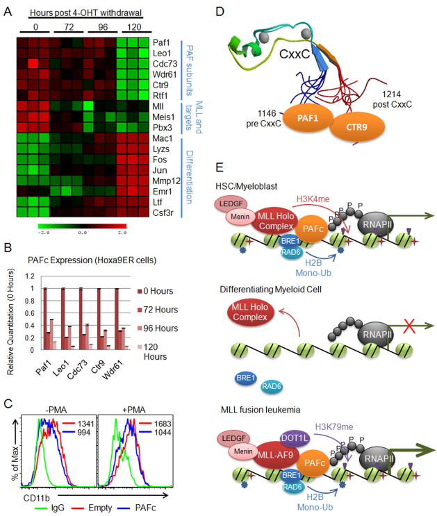
Figure 8. PAFc is down regulated during differentiation of hematopoietic cells.
A) Heat map generated from expression array data collected from differentiation of the Hoxa9ER cell line. Data is shown in triplicate for each time point following tamoxifen withdrawal. Labels indicate components of the PAF complex, MLL and MLL targets, and genes associated with myeloid cell differentiation. B) Expression of PAF components were verified using qPCR by separately differentiating the Hoxa9ER cell line by tamoxifen withdrawal. Time points indicate hours post tamoxifen withdrawal that RNA was collected. Expression for each component is shown relative to 0 hours. Error bars indicate +/− SD. C) THP-1 cells were transfected with an equal mixture of PAFc expression vectors or empty vectors along with a GFP vector at a 5:1 ratio (PAFc/Empty:GFP). Half were treated with PMA to induce differentiation. Surface expression of CD11b was monitored by FACS in the GFP positive gated cell population to track differentiation. Mean fluorescence values are shown for each sample. D) Structure of the CxxC domain and flanking sequences (PDB code: 2JYI). Solution structure of MLL CxxC region shows the flanking sequences of the CxxC domain brought into close juxtaposition creating a binding surface for both PAF1 and CTR9 of the PAF complex. E) Model for myeloid cell differentiation showing (top) PAFc dependent recruitment of MLL to target genes in hematopoietic progenitors (HSC/myeloblasts). PAFc and MLL recruitment promotes H2B mono-ubiquitination and H3K4 and H3K79 methylation resulting in transcriptional activation. (Middle) More differentiated myeloid cells down regulate PAFc expression resulting in decreased recruitment and transactivation by MLL. (Bottom) In leukemic cells harboring MLL fusion proteins the fusion protein synergizes with PAFc resulting in robust transcriptional activation of target genes. This is associated with increased H2B mono-ubiquitination and histone H3K79 methylation by DOT1L recruited through the MLL fusion partner. (See also Figure S6)

