
Figure 5.
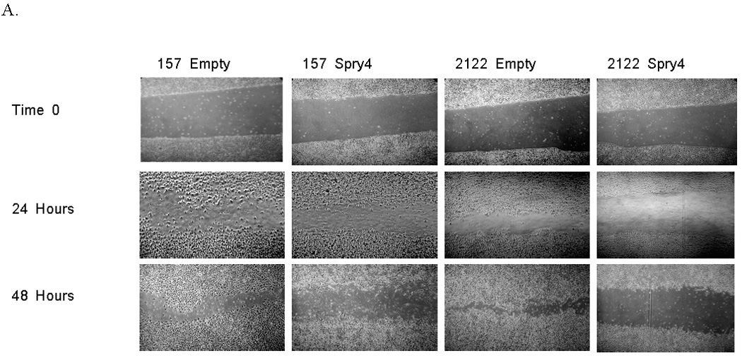

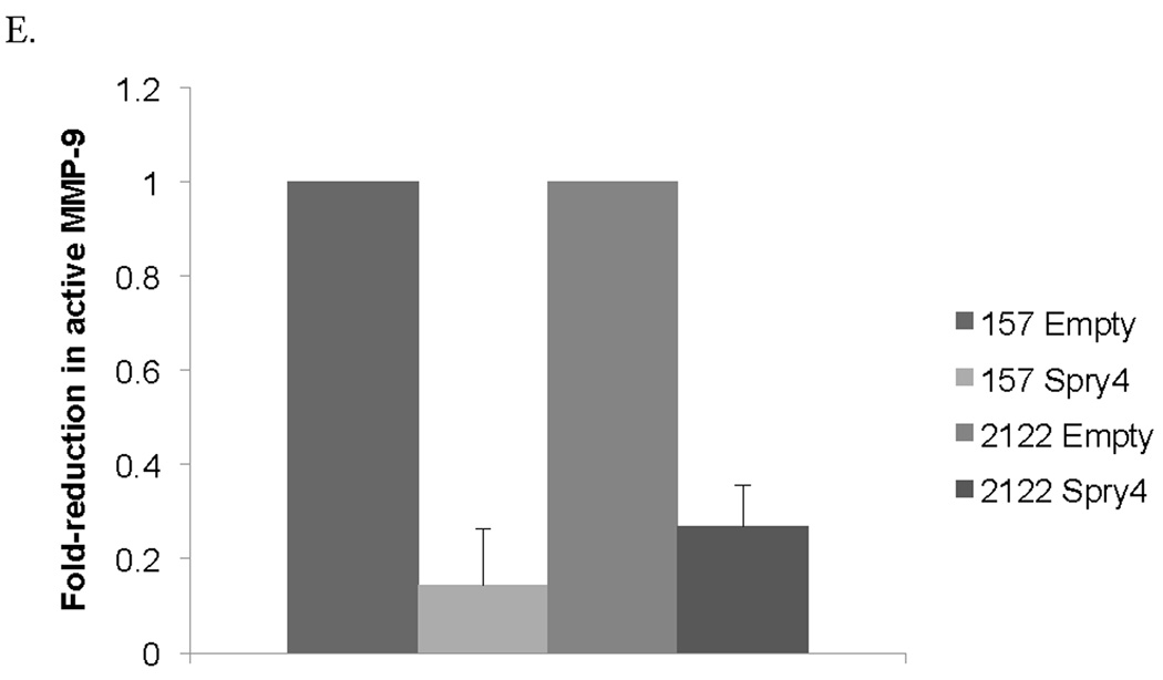
Re-expression of Spry4 inhibits migration and invasion in NSCLC cells. (A) A 3mm space was created across the diameter of plates of H157 and H2122 cells stably expressing Spry4 or an empty vector control and migration was recorded at 24 and 48 hours. Results are representative of triplicate experiments. (B) The 3D invasion assay uses a layer of collagen beneath a layer of matrigel and media containing the cells of interest. 5,000 cells from H157 and H2122 stably expressing Spry4 or an empty vector control were seeded in triplicate in media with 4% matrigel on top of a 1:1 matrigel and collagen base layer. Cell invasion was recorded on day 5. (C) Expression levels of CD82 and TIMP1 were assessed by QPCR in H157 and H2122 cell lines stably expressing Spry4 or an empty vector control. Results are the average of triplicate experiments normalized to GAPDH. (D) Increased expression of CD82 was confirmed by western blot analysis of cell lysates from H157 and H2122 cell lines stably expressing Spry4 compared to an empty vector control. Loading control is β-actin. (e) H157 cells stably expressing Spry4, H2122 cells stably expressing Spry4, and corresponding empty vector controls were seeded in a 96-well plate and analyzed with the MMP-9 Human Biotrak Assay. Absorbance was measured and a standard curve was used to calculate the amount of active MMP-9 in the samples. Results are the average of triplicate experiments with standard error.