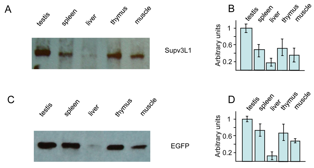
Figure 2.
Expression levels of Supv3L1 and EGFP reporter as assayed by Western blotting. (A) Tissue extracts were prepared from 10 weeks old wild-type mouse and analyzed using rabbit antibody against mouse Supv3L1. (B) Quantification of the data shown in (A) after correcting for loading variations using GAPDH as an internal standard. The results represent an average of three gels and are plotted in arbitrary units/mg of the protein extract. (C) Western blotting performed using tissue extracts obtained from 10 weeks old targeted (Supv3L1tm6Jkl/+) animal and rabbit antibodies against EGFP. (D) Quantification of the data shown in (C) representing an average of three gels.

