Figure 4.
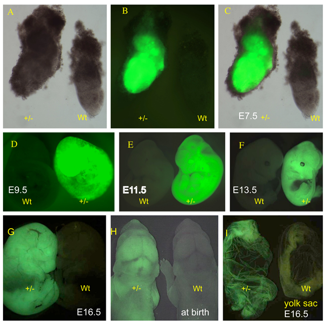
The EGFP reporter expression in postimplantation embryos. (A) Supv3L1tm6Jkl/+ embryo at E7.5 (+/−) and wild-type embryo (wt) photographed in the bright field. (B) EGFP emission of the same embryos shown in (A). (C) Merged images of (A) and (B). (D) EGFP expression in wild-type (wt) and Supv3L1tm6Jkl/+ (+/−) embryos at E9.5. (E) Fluorescent light emission in wild-type (wt) and Supv3L1tm6Jkl/+ (+/−) embryos at E11.5. (F) Wild-type (wt) and Supv3L1tm6Jkl/+ (+/−) embryos showing EGFP expression at E13.5. Note rather weak signal in the area of the developing liver. (G) The EGFP reporter expression at E16.5. (H) The EGFP reporter expression at birth. (I) Yolk sac from Supv3L1tm6Jkl/+ (+/−) and wild-type embryos at E16.5.

