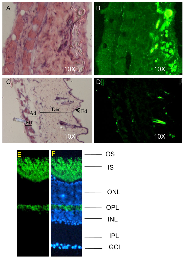
Fig. 6.
Detecting EGFP emission in tissue sections. (A) H&E stained section of the skin derived from Supv3L1tm6Jkl/+ animal. (B) Fluorescent light emission from the same section before H&E staining. Note that all cell layers including subcutaneous muscle fibers are positive. Bright fluorescent spots on the epidermal side of the skin are the hair shafts. (C) H&E stained section of the wild-type skin. Ad, adipose layer; Der, dermis; Ed, epidermis; Mf, subcutaneous muscle fibers. (D) Same section before H&E staining that shows no EGFP emission. Bright spots on the epidermal side of the crossection are the hair shafts that fluorize in the EGFP-independent manner. (E) Fluorescent light emission in mouse retina cryosections and (F) co-staining with hoechst (blue; nuclear stain). The signal is predominantly detected in photoreceptor inner segment (IS) and outer plexiform layer (OPL). OS: outer segment; INL: inner nuclear layer; IPL: inner plexiform layer; GCL: ganglion cell layer.

