
Figure 1.
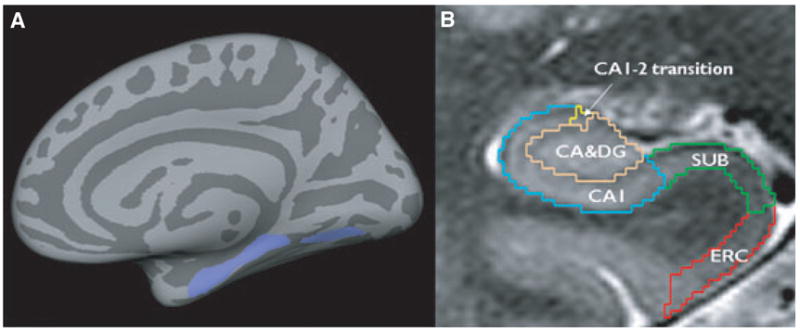
(A) Region of interest (ROI, lilac) containing parts of the ERC, parahippocampal, and lingual gyrus (ERC-paralingual ROI). Size and shape of the ROI were based on the region that showed significant correlations with the total ipsilateral thalamus volume in TLE-no and controls. (B) Manual marking scheme for hippocampal subfields. As it is not possible to identify individual hippocampal layers with 4T, the scheme was based on reliably recognizable anatomic landmarks, even though this resulted in a part of the prosubiculum and subiculum proper being counted toward the CA1 sector. ERC, entorhinal cortex; CA1-2, CA1-CA2 transition zone. Subfields were marked in the anterior part of the hippocampal body on a length of 1 cm, c.f. references in text for a detailed explanation of the marking scheme.
Epilepsia © ILAE